The Brain Just Got 25 Times More Detailed, and AI Did the Mapping
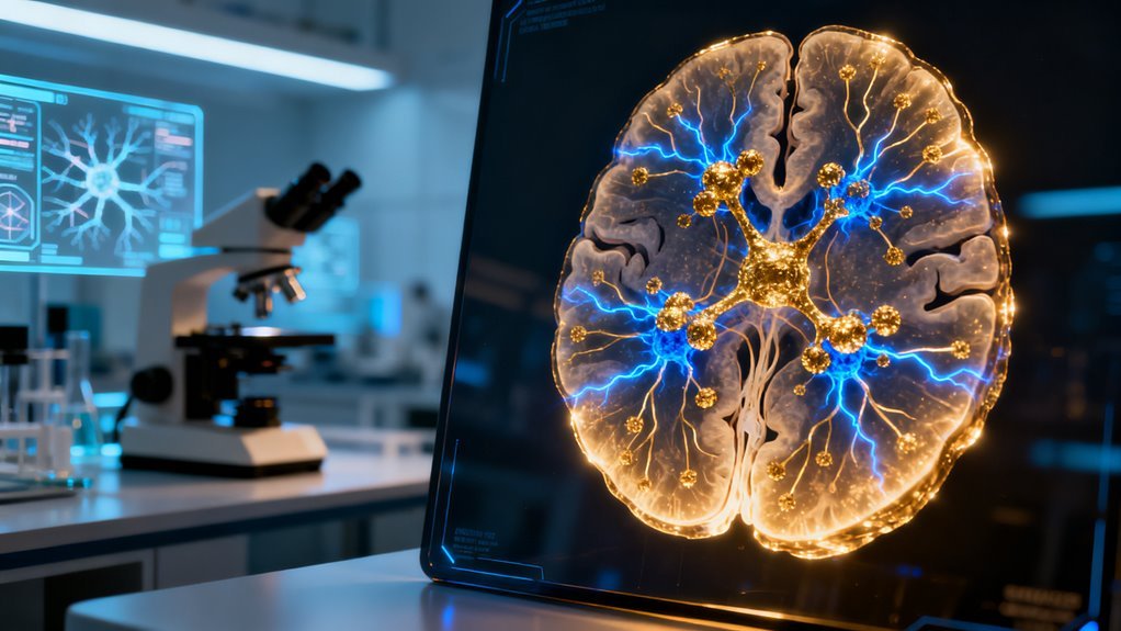
For decades, neuroscience operated with what amounted to a rough sketch of the brain. Fifty two mapped regions in the mouse brain served as the canonical reference, a framework that guided everything from drug development to our understanding of psychiatric disorders. That number just jumped to approximately 1,300. The tool responsible is CellTransformer, an AI system trained on single cell RNA profiles drawn from nine million neurons, and its findings don’t just refine the existing map. They expose structures that conventional imaging and molecular techniques couldn’t see at all.
What makes this worth paying attention to isn’t the sheer number of newly identified microregions, though that alone represents a staggering expansion. It’s where some of those regions turned up and what they appear to do.
Motivation Has a Geography We Didn’t Know Existed
Among the newly charted areas are microregions embedded within the nucleus accumbens and ventral tegmental area, two structures that neuroscience has studied intensively for decades because of their central role in reward, motivation, and dopamine signaling. Researchers thought they understood the architecture of these regions reasonably well. CellTransformer suggests otherwise.
The identification of distinct substructures within these areas carries immediate implications. Current models of reinforcement learning, both biological and computational, treat the reward circuitry as relatively uniform at the cellular level. If these microregions turn out to have functionally distinct roles in dopamine modulation, the existing frameworks need revision. That’s not a minor update. Reinforcement learning from human feedback, the technique underpinning alignment strategies at OpenAI, Anthropic, and others, draws conceptual inspiration from the same biological reward mechanisms now being revealed as far more granular than assumed.
This doesn’t mean RLHF is broken. But it does mean that our simplified models of how reward signals operate in biological brains were masking a layer of complexity that could eventually inform better computational analogs.
Why AI Found What Traditional Methods Missed
The key technical insight here is scale and pattern recognition operating simultaneously. Traditional single cell RNA sequencing could profile thousands, sometimes hundreds of thousands, of neurons. But identifying coherent spatial microregions within known structures requires analyzing millions of cells in parallel, detecting subtle expression gradients that would be invisible in smaller datasets or through manual annotation.
CellTransformer belongs to a growing class of foundation models built for biological data. The approach mirrors what has happened in language and vision. Train at sufficient scale on the right data, and emergent structure appears. Google DeepMind demonstrated a version of this logic with AlphaFold, which reshaped structural biology not by introducing fundamentally new physics but by processing protein sequences at a scale and depth that revealed patterns human researchers could not detect unaided.
The parallel matters because it signals a broader trend: AI is becoming the primary discovery instrument in the life sciences, not merely an assistive tool for hypothesis testing. When a model trained on nine million neurons identifies 1,248 regions that human researchers missed across decades of careful study, it raises a straightforward question about where the bottleneck in scientific discovery actually sits. Increasingly, the answer appears to be human perceptual and cognitive limits rather than data availability.
Precision Psychiatry Gets a New Foundation
The practical downstream impact concentrates most immediately in psychiatry and neuropharmacology. Depression, addiction, ADHD, and anhedonia all involve dysfunction in the reward and motivation circuits that CellTransformer has now subdivided into finer components. Current psychiatric medications are notoriously imprecise. SSRIs flood entire neural systems with serotonin. Stimulants broadly amplify dopamine. The field has long recognized that this blunt approach produces inconsistent outcomes, but lacked the anatomical resolution to do better.
If distinct microregions within the nucleus accumbens govern separable aspects of motivation and reward processing, targeted interventions become theoretically possible. That could mean more selective drug design, more precise deep brain stimulation protocols, or better biomarkers for diagnosing subtypes of conditions currently lumped under single diagnostic labels.
The timeline for clinical translation is long. We are talking years, likely a decade or more, before these mapped microregions yield approved therapies. But the mapping itself removes a fundamental obstacle. You cannot target what you cannot see.
What People Are Overlooking
The conversation around AI in neuroscience tends to focus on brain computer interfaces and companies like Neuralink. That emphasis is understandable given the consumer visibility, but it obscures a potentially larger story. Computational tools that reveal previously unknown brain architecture may matter more in the long run than devices that interface with the architecture we already knew about.
There is also a governance question that deserves attention. As AI driven brain mapping becomes more detailed and potentially applicable to human tissue, the intersection with neurorights legislation, still in its infancy globally, will become increasingly relevant. Chile passed the world’s first neurorights law in 2021. Others are watching. A 25 fold increase in brain cartographic resolution, even in a model organism, pushes these policy conversations forward whether regulators are ready or not.
The direction is clear. AI is not just accelerating neuroscience. It is redefining what neuroscience can observe. And when the instrument changes what’s visible, the science that follows tends to change everything else.
For decades, scientists have carved the human brain into roughly 52 distinct regions, a number that traces back to Korbinian Brodmann’s early twentieth century maps and has been refined only incrementally since. That number just jumped to approximately 1,300. A transformer architecture called CellTransformer, trained on single cell RNA profiles from 9 million neurons, has redrawn the map of the brain with a granularity that makes previous atlases look like sketching continents on a napkin. This is not a modest improvement. It is a twenty five fold expansion of what neuroscience considers charted territory, and it arrived not through painstaking manual dissection but through the same class of AI architecture that powers large language models.
The significance here extends well beyond an academic exercise in neuroanatomy. What CellTransformer accomplished is pattern recognition at a scale and depth that human researchers simply cannot replicate manually, even with unlimited time. The model treated transcriptomic data the way GPT treats language: it learned the underlying grammar of how cells relate to one another, identified clusters that had never been formally distinguished, and did so in hours rather than years. That speed matters. It signals that AI is no longer just accelerating existing scientific workflows. It is revealing structures and relationships that were genuinely invisible to prior methods, much like the PULSE program aims to identify key health insights for public health applications.
Why the Timing Is Not Accidental
This breakthrough sits at the intersection of two converging capabilities that only recently matured. First, single cell RNA sequencing has dropped in cost and increased in throughput dramatically over the past five years, making datasets of 9 million neurons feasible rather than aspirational.
Second, transformer architectures have proven their versatility far beyond text. We have seen them applied to protein folding with AlphaFold, molecular design, weather prediction, and genomics. CellTransformer is the logical next step in that migration, but the results are striking because the domain itself, neuroanatomy, had remained stubbornly resistant to high resolution automation.
Google DeepMind’s work on AlphaFold reshaped structural biology almost overnight. CellTransformer may do something analogous for neuroscience, though the downstream consequences could prove even more complex. Understanding where proteins fold is one thing. Understanding the fine grained architecture of the organ that produces consciousness, motivation, memory, and disease is quite another. The research team behind this work included experts from the University of California, San Francisco, and the Allen Institute for Brain Science, institutions with deep expertise in both computational methods and neuroanatomy.
What the New Map Actually Reveals
The expanded atlas is not just a finer slicing of previously known regions. It exposes microregions that had no prior classification at all. Among the most significant findings are new subdivisions within structures central to motivation and reward, areas that matter enormously for understanding addiction, depression, ADHD, and a range of psychiatric conditions where existing treatments remain blunt instruments.
The nucleus accumbens, long understood as a core hub for wanting and reward seeking behavior, now appears to contain internal architecture that previous methods collapsed into a single functional unit. The ventral tegmental area, which supplies dopamine to much of the brain’s motivation circuitry, shows similar internal complexity.
Additional microregions in the ventral pallidum, where neurons encode motivational states by responding with positive signals to rewards and negative signals to punishments, have surfaced with specificity that was previously unavailable. Cortical contributions from orbitofrontal, anterior cingulate, medial prefrontal, and insular regions also gain new detail in the atlas.
These areas integrate affective salience with motor plans and utility computations, meaning they sit at the crossroads of feeling, deciding, and acting. Mapping them at cellular resolution opens the door to understanding not just what these regions do in broad strokes, but precisely which cell populations within them are responsible for specific computational roles.
Dopamine Neurons as Temporal Clocks
One finding deserves particular attention because it challenges a framework that has dominated computational neuroscience for more than two decades. Machine learning analysis of ventral tegmental area firing patterns reveals that dopamine neurons do not merely signal reward prediction errors, the mismatch between expected and received rewards that forms the backbone of reinforcement learning theory.
They also function as temporal clocks, encoding both the expected magnitude of future rewards and the precise timing of when those rewards should arrive. This distinction matters for AI as much as it does for neuroscience. Reinforcement learning algorithms inspired by dopamine signaling power everything from robotics to recommendation systems.
If the biological system is performing richer computations than the simplified models that AI researchers borrowed from neuroscience, then there may be room for fundamentally improved RL architectures that incorporate temporal prediction more deeply. The feedback loop between neuroscience and AI has always been bidirectional, and this finding could send new inspiration flowing back toward machine learning.
Dopamine release downstream operates as a neuromodulatory signal that triggers actions in anticipation of positive stimuli. The neurons are not just reacting to what happened. They are forecasting what will happen and when, anchoring motivated behavior to time expectations. That is a far more sophisticated computation than classical models assumed, and the AI analysis is what made it visible.
Intrinsic Motivation Gets a Biological Address
Perhaps the most provocative result for anyone thinking about artificial general intelligence or advanced AI systems is the identification of specialized dopaminergic neuron types that underpin intrinsic motivation. The atlas distinguishes value coding subpopulations, which signal expected reward value, from salience coding subpopulations, which fire in response to motivationally significant events regardless of whether they are positive or negative.
The anterior insula receives direct input from both types and appears to contextualize motivational signals for adaptive decision making. These circuits rely on evolutionarily ancient dopaminergic systems, which suggests that curiosity driven exploration and mastery seeking behaviors are not recent cognitive luxuries. They are deep biological imperatives, hardwired into some of the oldest neural architecture we carry.
For the AI research community, this finding raises an interesting question. Current approaches to intrinsic motivation in artificial agents, such as curiosity driven exploration in reinforcement learning, are loosely inspired by neuroscience but operate on simplified assumptions.
Having a detailed cellular map of how biological intrinsic motivation actually works could inform the next generation of architectures designed to produce genuinely exploratory, self directed behavior in machines.
Who Benefits and What Changes
The immediate beneficiaries are neuroscience researchers who now have a reference atlas of unprecedented detail. But the downstream effects ripple outward considerably.
Pharmaceutical companies developing treatments for psychiatric and neurological conditions gain a much finer target map. Today’s psychiatric drugs are notoriously imprecise because they modulate neurotransmitter systems across the entire brain rather than targeting specific microregions.
With 1,300 distinct cellular domains identified, drug development can begin moving toward precision psychiatry in a way that was previously aspirational. If a specific microregion within the nucleus accumbens is implicated in compulsive reward seeking while a neighboring microregion handles adaptive motivation, the therapeutic implications are enormous.
Neurotech companies working on brain computer interfaces also stand to benefit. Neuralink, Synchron, Precision Neuroscience, and others are building devices that read from and write to neural tissue. A twenty five fold increase in the resolution of brain maps directly improves the precision with which these devices can be designed, placed, and calibrated.
Knowing exactly which cell populations occupy a target region changes the engineering calculus for electrode design and stimulation protocols. For AI developers, the enriched understanding of biological motivation circuits offers new computational metaphors to explore.
The history of AI is littered with productive borrowings from neuroscience, from perceptrons to attention mechanisms to reward prediction errors. A detailed atlas of how the brain organizes motivation at the cellular level is fresh material for architectures that aim to produce adaptive, goal directed behavior.
What People Are Overlooking
The conversation around this work will likely focus on the neuroscience findings themselves, and that is understandable. But the methodological story deserves equal weight. CellTransformer demonstrates that transformer architectures can serve as discovery engines in domains with complex, high dimensional biological data.
The model did not just confirm what was already known. It found structures that experts had not identified. That capability, applied systematically, could transform cell biology, developmental biology, immunology, and oncology with similar force.
There is also a governance question that nobody is yet asking loudly enough. As AI powered atlases become the reference standard for neuroscience, who controls the models, the training data, and the interpretive frameworks?
If a single model becomes the canonical tool for brain mapping, the biases embedded in its training data or architecture could shape an entire generation of neuroscience research. The scientific community will need to develop validation standards and replication protocols specific to AI generated biological atlases, a challenge that has no established playbook.
Looking Forward
The pace at which AI is revealing biological complexity that eluded traditional methods is accelerating. AlphaFold mapped protein structures. Large language models are decoding gene regulation. Now CellTransformer has redrawn the brain’s map at a resolution that would have taken human researchers decades to achieve, if they could have achieved it at all.
What comes next is the integration phase. Linking these 1,300 cellular domains to specific functions, behaviors, and disease states is a project that will occupy neuroscience for years. But the foundation is now in place, and it was built by the same class of architecture that currently dominates commercial AI.
The boundary between artificial intelligence as a product and artificial intelligence as a scientific instrument is dissolving rapidly, and this work is one of the clearest examples yet of what that convergence produces.