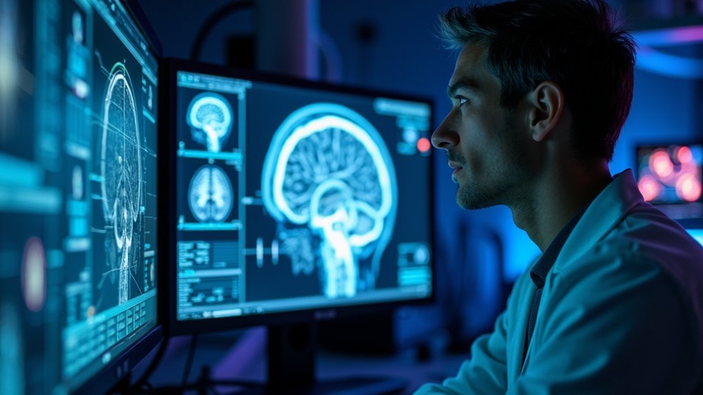

Artificial intelligence is quietly changing how depression is studied, not by chasing buzzwords but by giving researchers sharper instruments to see what is happening in the brain and behavior. Instead of relying only on symptom scales and clinical hunches, teams are now using machine learning to read patterns in brain signals that were previously invisible, with the goal of building more precise and reliable models of major depressive disorder. AI digital twins could enhance the understanding of individual health trajectories in patients with depression.
How we got here
For most of modern psychiatry, depression has been defined by what people report and how clinicians interpret those reports. Rating scales, clinical interviews, and broad diagnostic categories have driven research and treatment decisions since the late twentieth century. This approach has helped standardize care but treats depression as a single label applied to people with very different experiences, from severe anhedonia to agitated anxiety, often bundled together under the same code.
Depression has long been reduced to shared checklists, obscuring wildly divergent lived experiences
Neuroimaging changed the conversation by revealing that depression is associated with alterations in prefrontal control circuits, limbic emotion networks, and reward pathways, but early findings were often inconsistent and hard to translate into practice. Different studies focused on single brain regions or used small samples, which made it difficult to separate signal from noise. The rise of machine learning offered a way to move past simple region-by-region comparisons and instead analyze whole brain connectivity and complex signal patterns at once.
AI models that read the depressed brain
Recent work with deep learning and graph-based models illustrates how far this field has come. The Brain Augmented Decorrelated Network, known as BrainADNet, uses a graph convolutional architecture to learn from resting state brain signals while also integrating demographic information such as age, sex, and education. By encouraging the model to extract non-redundant features from multi-layer brain connectivity graphs, BrainADNet improves classification of individuals with major depressive disorder compared with conventional machine learning approaches that rely on simpler feature sets. In head-to-head studies, BrainADNet outperformed existing methods in identifying major depressive disorder, revealing distinct connectivity signatures that track with depression severity and stage.
The practical consequence is that multivariate brain networks carry clinically relevant information even when single region measures appear inconclusive. Instead of asking whether one prefrontal area is more or less active, models like BrainADNet examine how multiple regions talk to each other across time and how those communication patterns differ between depressed and non-depressed groups. This shift from isolated signals to network-level analysis reflects a broader change in neuroscience toward viewing psychiatric disorders as circuit problems rather than purely chemical imbalances.
From fixed labels to brain-based dimensions
Another important step has been moving beyond strict categorical diagnosis. Traditional systems assign people to depression or no depression, but clinicians know that two patients with the same diagnosis can share few symptoms. AI-based dimensional neuroimaging frameworks take advantage of continuous brain-based axes, mapping symptom intensity onto distributed neural circuits rather than collapsing everything into a single label.
In these approaches, features such as anhedonia, anxiety, and cognitive impairment are treated as graded dimensions that appear in patterns of activation and connectivity across large-scale networks. Machine learning models identify clusters of individuals who share similar circuit-level abnormalities even when their symptom profiles overlap only partially. This perspective reframes depression as a family of related brain network disorders rather than one homogeneous condition and provides a conceptual foundation for precision psychiatry. It is a promising direction, but it also demands rigorous validation to ensure that these data-driven subgroups correspond to stable and clinically meaningful entities.
Biotypes and circuit-informed treatment
One of the most widely discussed developments is the identification of biological subtypes, or biotypes, of depression using brain imaging combined with machine learning. A recent study using functional imaging and clustering algorithms reported six distinct biotypes of depression and anxiety, each defined by a unique pattern of activity across key mood and cognitive control regions. Some biotypes showed disrupted communication between prefrontal control regions and limbic structures, others showed thalamic hyperconnectivity, and still others involved pronounced changes in reward circuitry.
These patterns were not just academic curiosities. The study linked several biotypes to differential responses to treatments, including specific antidepressant medications, neuromodulation techniques, and psychotherapy modalities, suggesting that certain circuit profiles are more likely to respond to particular interventions. In research settings, this opens the door to circuit-informed treatment selection, moving beyond trial-and-error prescribing toward strategies guided by objective neurobiological signatures. The next challenge is to test these biotype-based recommendations prospectively, track long-term outcomes, and see whether imaging-derived labels retain their predictive value outside controlled cohorts.
EEG and fNIRS give portable windows into depression
Not all brain-based AI work depends on high-end scanners. Electroencephalography and functional near-infrared spectroscopy offer more accessible ways to capture depression-related brain activity and have become fertile ground for machine learning research. EEG provides millisecond-level timing of neural oscillations, which allows models to detect spectral and connectivity features associated with psychomotor slowing, rumination, and other depressive phenomena.
A hybrid EEG and fNIRS study illustrates the power of combining modalities. Using machine learning, researchers reported that classification accuracy was about eighty-two percent with EEG features alone and rose to nearly ninety-three percent when EEG was combined with fNIRS measures of prefrontal oxygenation. The most informative features included local efficiency in delta band brain networks, hemispheric asymmetry in theta band activity, and sample entropy measures of brain oxygenation, pointing to coordinated electrical and hemodynamic changes in depression.
Functional near-infrared spectroscopy on its own has also shown promise. Studies using verbal fluency tasks and prefrontal fNIRS recordings have reported significant differences in hemodynamic responses between people with major depressive disorder and healthy controls, with machine learning classifiers achieving respectable performance in distinguishing the two groups. Feature importance analyses consistently highlight dorsolateral and medial prefrontal regions as key contributors, reinforcing the view that altered frontal control processes are central in depression.
More recently, simultaneous resting state EEG and fNIRS experiments have been used to study neurovascular coupling in depression, revealing coordinated changes in electrical activity and blood oxygen dynamics that may serve as multimodal biomarkers of both depressive states and recovery over time. This line of work is particularly interesting because EEG and fNIRS can be made wearable and relatively inexpensive, hinting at future scenarios where objective brain measurements might be collected in outpatient clinics or even at home.
AI for predicting treatment response
Beyond diagnosis, AI models are increasingly being trained to predict how people will respond to treatment. In a study combining fNIRS with machine learning, changes in total hemoglobin during a prefrontal task were significantly correlated with reductions in depression scores after six months, and a naive Bayes model using only fNIRS data achieved balanced accuracies around seventy percent for predicting treatment response. Another analysis linked baseline prefrontal activity patterns, especially in dorsolateral prefrontal cortex, to subsequent improvement in Hamilton Depression Rating Scale scores, suggesting that fNIRS signals might help identify who is more likely to benefit from a given intervention.
Related work has used gradient boosted decision trees and other algorithms applied to multi-feature fNIRS patterns to classify major depressive disorder with high sensitivity and area under the curve, again pointing to the frontopolar and dorsolateral prefrontal regions as particularly informative. When clinical data are combined with spectroscopy features, some studies find incremental improvements in diagnostic accuracy, although results are mixed and underscore the need for careful model evaluation across diverse populations.
There is also a growing body of research focusing on enhanced EEG-based classification of major depression through advanced feature extraction and domain adaptation techniques, aiming to make models more robust to differences in recording conditions and patient characteristics. Taken together, these efforts show that AI is not only being used to identify who is currently depressed but increasingly to forecast who will improve and with which treatments.
Opportunities, limits, and real-world risks
The opportunities are clear. If robust brain-based biomarkers can be validated, clinicians could move toward more personalized care, matching patients to therapies that fit their circuit profile rather than cycling through multiple medications and interventions in search of something that works. Portable modalities like EEG and fNIRS make it plausible that such tools could extend beyond academic centers into community settings, provided that devices and workflows are simplified enough for routine use.
Yet the limitations are equally important. Despite encouraging accuracies and compelling brain maps, clinically actionable imaging biomarkers that reliably distinguish depression from closely related conditions such as anxiety disorders or bipolar depression remain unvalidated. Many studies involve modest sample sizes, single sites, and selective inclusion criteria, which raise concerns about generalizability. Cross-validation can overestimate performance when data are not carefully separated, and models trained on one population may perform poorly in another.
There are also ethical questions. Brain-based AI tools could unintentionally reinforce biases if training data underrepresent certain demographic groups or if models are applied without careful calibration to local populations. Overreliance on algorithmic outputs might sideline lived experience and clinical judgment, especially if tools are marketed as objective solutions without transparent discussion of their uncertainty. Privacy and data security are ongoing concerns in any system that records sensitive neural and behavioral information.
What this means for the future of depression care
Looking across these developments, a few themes stand out. First, AI is helping shift depression research from symptom checklists to circuit-level models, integrating structural and functional brain data with clinical and demographic information to uncover patterns that were previously inaccessible.
Second, multimodal signal analysis using EEG, fNIRS, and imaging is revealing consistent involvement of prefrontal control regions, limbic networks, and reward circuits, while at the same time exposing meaningful heterogeneity in how these systems are disrupted across individuals.
Third, early evidence that biotypes and brain-based features can predict treatment response hints at a future in which precision psychiatry is not just a slogan but a practical framework for guiding decisions about medication, psychotherapy, and neuromodulation. However, that future will depend on large-scale replication, careful external validation, and thoughtful integration into clinical workflows rather than standalone technologies.
In the near term, the most realistic path forward is for AI-enhanced brain measures to serve as auxiliary tools that complement clinical assessment, helping to clarify ambiguous cases, flag treatment resistance earlier, and generate testable hypotheses about which circuits to target in new interventions. Over time, as evidence accumulates and standards mature, some of these tools may earn their place as routine components of depression care. The signal is promising, but the field will be judged not by how sophisticated the models look today, but by whether they ultimately improve outcomes for people living with depression.
Conclusion
Why AI brain mapping for depression matters now
Depression is common, complex, and still very hard to diagnose and treat precisely. Standard clinical assessments rely on what people report about their mood, sleep, appetite, and functioning, which can be subjective and variable over time. At the same time, modern brain imaging produces enormous amounts of data that are almost impossible for humans to parse fully. AI systems are now starting to bridge that gap, searching through brain signals to uncover subtle patterns that correlate with depressive symptoms and treatment response.
This matters now for two reasons. First, clinical demand is rising while specialist resources remain limited. Second, the field has reached a point where AI is not just labeling scans but extracting candidate biological signatures of depression that could reshape how we diagnose, monitor, and treat the disorder.
How we got here: from brain scans to algorithmic signatures
For decades, researchers have known that depression is associated with changes in specific brain regions and networks. Functional MRI studies repeatedly highlighted areas such as the amygdala, anterior cingulate cortex, and prefrontal cortex in emotional regulation and negative bias. Structural and connectivity studies added evidence that networks linking limbic and frontal regions are often disrupted in major depressive disorder.
The challenge was scale. A single neuroimaging session can contain millions of data points across space and time. Early work used relatively simple statistical models that looked at predefined regions of interest or averaged activity across networks. These approaches established important associations but were limited in their ability to capture complex, distributed patterns.
Machine learning methods began to change this in a meaningful way. Reviews of depression imaging have catalogued studies where algorithms classified patients versus healthy controls based on whole brain connectivity, sometimes reaching very high sensitivity by focusing on network level features across default mode, affective, visual, and cerebellar systems. In parallel, electroencephalography and other modalities provided time resolved signatures that learning algorithms could exploit.
Today, deep learning and graph based models are being trained directly on resting state brain signals, sometimes combining imaging with demographic and behavioral data. These systems do not just ask whether a brain looks “depressed” overall. They search for fine grained connectivity motifs and activation patterns that track symptom severity, subtypes, and treatment outcomes.
What current AI studies are actually finding in the brain
Recent research illustrates how far this approach has progressed and where it is still provisional.
One line of work focuses on brain connectivity patterns. At the Indian Institute of Technology Delhi, researchers built a deep learning framework called Brain Augmented Decorrelated Network that uses graph convolutional techniques to analyze resting state imaging signals together with demographic attributes such as age, gender, and education. The model improved diagnostic accuracy across multiple stages of depression and highlighted specific brain regions whose connectivity patterns were most influential for classification, including differences between male and female patients. This kind of analysis begins to map depression onto distinct network signatures rather than a single generic profile.
Another study presented at the Society for Neuroscience used deep learning on magnetic resonance imaging data from 1,162 participants, including 334 people with depression. By scanning thousands of connections, the algorithms identified 83 abnormal connectivity patterns correlated with depressive status. Many of these involved motor networks and the thalamus, a region central to emotional regulation. The fact that electrical stimulation of the thalamus has been linked to improvement in depressive symptoms adds biological plausibility to these algorithmic findings.
Meta analytic work reinforces the idea that depression is best understood through multi modal patterns. A recent systematic review and meta analysis examined AI assisted screening methods that combine voice, facial expression, eye movements, and EEG or imaging data. It found that multi modal fusion systems can distinguish depressed from non depressed individuals with diagnostic accuracy approaching 96 percent in some configurations, with strong sensitivity and specificity. These systems detect features such as slowed speech, increased negative facial expressions, biased attention toward negative stimuli, and abnormal activation in regions like dorsolateral prefrontal cortex, anterior cingulate cortex, orbitofrontal cortex, and amygdala.
Beyond diagnosis, AI is being used to predict symptom change under treatment. In one study, algorithms trained on brainwave measurements in depressed individuals were able to predict which specific symptoms would improve with antidepressant treatment. The strongest performance was seen for a subset of symptoms including insight and weight change. This kind of symptom level prediction begins to move from “Does treatment work” to “Which aspects of depression will this treatment help for this particular person.”
Taken together, these studies show that AI can detect meaningful brain patterns linked to depression status and severity, and sometimes to clinical trajectory. At the same time, they remain primarily research tools, not fully validated clinical instruments.
Why this could reshape clinical practice
If these systems continue to improve and are validated across diverse populations, the impact on clinical practice could be substantial.
More precise and earlier diagnosis
Current diagnosis of major depression relies on symptom checklists and clinical interviews. Two people can receive the same diagnosis while having very different underlying biology and clinical trajectories. AI assisted imaging and multi modal data could help clinicians:
- Identify individuals whose brain connectivity patterns and behavioral signals suggest high risk of progressing to more severe or chronic depression even when symptoms are still mild.
- Distinguish depression from other conditions that share features such as anxiety, cognitive impairment, or fatigue, by focusing on pattern combinations that are more specific to depressive pathology.
- Recognize subtypes based on network level profiles, similar to work that has already defined distinct neurophysiological biotypes using functional connectivity patterns in limbic and frontostriatal circuits.
Matching treatments to brain and behavior
AI driven pattern recognition could also support more tailored treatment strategies. For example:
- If a model indicates that certain emotional regulation networks are particularly disrupted, a clinician might prioritize psychotherapies targeting cognitive reappraisal or behavioral activation, or neuromodulation techniques aimed at those circuits.
- When brainwave based algorithms predict that only specific symptoms are likely to respond to a given antidepressant, clinicians could adjust medication plans and set more realistic expectations with patients.
- Imaging and multi modal signals can be tracked over time, allowing models to flag when a patient is not responding as expected even before clinical symptoms fully reveal that lack of progress, which could prompt earlier treatment adjustments.
Informing prevention strategies
On the prevention side, the same technologies could help identify neural and behavioral markers of resilience as well as vulnerability. By contrasting individuals with similar life stress exposures but different outcome trajectories, AI systems can search for protective connectivity and activation patterns. Recognizing these signatures could guide interventions that reinforce more adaptive processing of emotion and stress.
The wider societal context: AI, mental health, and everyday use
There is another dimension that deserves attention. While AI is being used in laboratories and clinics to analyze depression, everyday use of generative AI tools appears to correlate with depressive symptoms in the general population. A large survey of more than twenty thousand adults in the United States found that daily use of generative AI was associated with significantly higher odds of reporting at least moderate depression, even after adjusting for sociodemographic factors.
In that study, people who used AI each day had roughly thirty percent higher odds of moderate depression compared with non users, with the association particularly strong among adults aged twenty five to sixty four. The magnitude of these associations was modest but consistent across depressive, anxiety, and irritability symptoms. The findings do not prove causation, but they underscore that AI currently has two faces in mental health. In research settings it is a powerful analytic tool. In everyday life its use is intertwined with habits and contexts that may not always be beneficial.
For technology companies and health systems, this duality is important. AI designed to support mental health must be evaluated not only for accuracy on lab datasets, but also for its impact on how people feel and behave when they interact with these tools routinely.
Limits, risks, and ethical questions
The scientific results are promising, yet they come with important caveats and ethical concerns that need to be addressed openly.
Correlation versus causation
Most current AI studies in depression are cross sectional or observational. They show that certain patterns of brain connectivity, activation, or behavior are associated with depressive states, but they do not establish whether those patterns cause depression, result from it, or reflect compensatory mechanisms. Even when AI models predict treatment response, they may be capturing markers of general health, social support, or other hidden variables. Robust causal inference will require prospective designs, interventions guided by AI discovered signatures, and mechanistic studies that link network changes to specific psychological processes.
Data diversity and generalizability
Many imaging datasets used in these studies are limited in size, geography, and demographic diversity. Some deep learning models have been trained on hundreds rather than tens of thousands of patients, often from single countries or health systems. That raises questions about how well their discovered patterns generalize to populations with different genetic backgrounds, social contexts, and comorbidities. Demographic attributes can be integrated into models, but that does not fully solve the underlying data imbalance.
Interpretability and clinical trust
AI models can outline intricate patterns of activity and connectivity, but they do not automatically explain what those patterns mean for cognition, emotion, and daily functioning. As researcher Terrence Sejnowski has pointed out, AI can reveal the movements and correlations in brain data, yet humans still must interpret how those micro details map to memory, thought, perception, and subjective experience. Clinicians are rightly cautious about acting on opaque signals. Tools that highlight specific regions and connections along with probabilistic predictions, and that can be validated against known neurobiology, are more likely to gain trust.
Privacy, consent, and misuse
Brain imaging and multi modal behavioral data are intensely personal. The prospect of models that can infer mental health status from voice, facial expression, or eye movements raises serious privacy concerns. The same algorithms that screen for depression might be repurposed for surveillance, employment screening, or insurance decisions if guardrails are not established. Ethical deployment will require strict data governance, meaningful consent, limits on secondary use, and oversight mechanisms that involve clinicians, patients, and ethicists.
What to watch next
Looking ahead, several developments will determine whether AI based brain pattern analysis for depression moves from promising research to trustworthy clinical practice.
- Larger, more diverse multi site datasets that combine imaging, electrophysiology, behavior, and long term clinical outcomes will be crucial. These will help test whether current signatures hold up across populations and care settings.
- Prospective trials where treatment choices are guided in part by AI derived brain and behavior patterns can show whether these tools improve outcomes compared with standard care.
- Efforts to integrate interpretability into model design, for example through explicit mapping of learned features onto known networks and cognitive processes, can help clinicians understand and critique algorithmic suggestions.
- Close collaboration between technologists, neuroscientists, and front line clinicians will be essential. Many promising systems fail not because the algorithms are weak, but because they do not fit into real clinical workflows or address the practical questions that matter to patients and practitioners.
At its best, this line of research offers a more nuanced map of neural vulnerability and resilience in depression, moving beyond checklists toward biologically informed care. AI systems are turning vast imaging and multi modal datasets into signals that might refine diagnosis, support personalized interventions, and inform targeted prevention strategies. Yet their long term impact will depend on rigorous validation, careful attention to ethics, and ongoing collaboration to ensure that algorithmic insights genuinely help patients across the full diversity of real world contexts. reddit