AI Is Learning to Spot Blood Cancer in Routine Lab Work, and the Implications Go Far Beyond Oncology
For decades, catching blood cancers early has been a matter of luck as much as medicine. A patient visits a doctor for fatigue or a routine physical, blood gets drawn, and somewhere in the results a subtle anomaly catches a hematologist’s eye. Or it doesn’t. The window between a cancer’s first detectable trace in the bloodstream and the moment it produces obvious symptoms can stretch for months, sometimes years. That gap has always been one of oncology’s cruelest features.
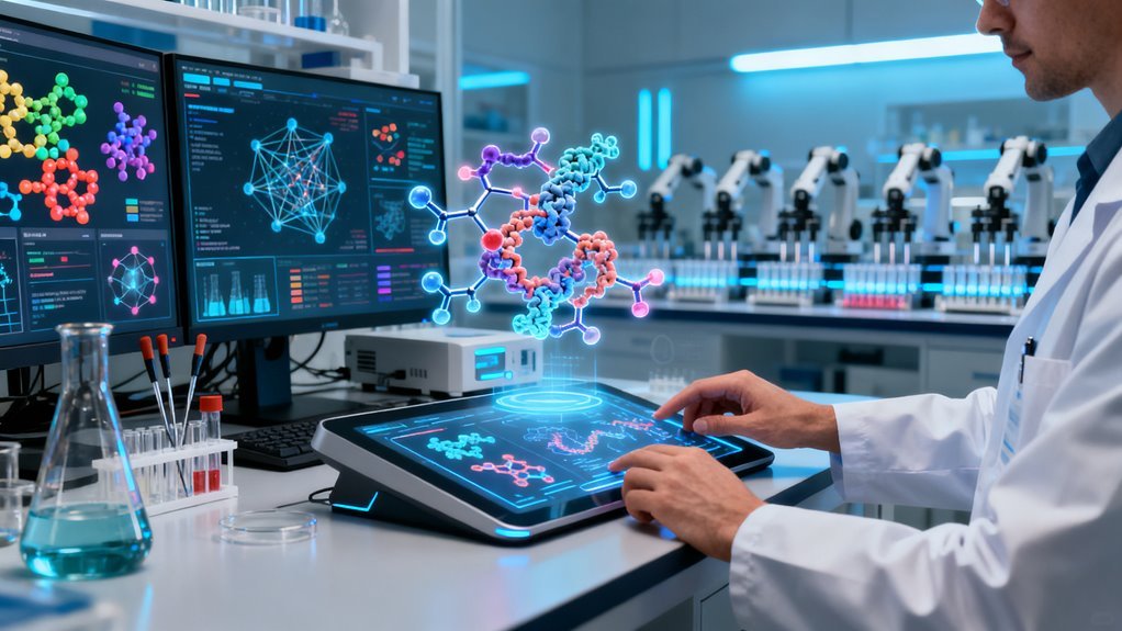
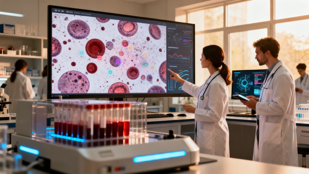
Now a growing body of clinical evidence suggests AI models can reliably close that gap by analyzing the same routine blood work patients already receive, flagging suspicious patterns long before a human reviewer would notice them. Deep learning systems trained on peripheral blood smear images are posting detection accuracies north of 97 percent. Machine learning algorithms running against standard hematology analyzer output reach roughly 88 percent sensitivity for acute leukemias. These are not theoretical benchmarks from a controlled dataset. Multiple hospital systems are beginning to integrate these tools into live diagnostic workflows.
The significance extends well beyond hematology. What is happening in blood cancer detection represents one of the clearest demonstrations yet of a pattern that will reshape medicine over the next decade: AI models finding clinical signal in data that already exists, collected through tests that are already standard, without requiring new hardware, new sample types, or new patient behavior.
Why This Breakthrough Follows a Predictable Trajectory
The technical pieces behind this development have been falling into place for several years. Computer vision models capable of classifying cell morphology from blood smear images matured around 2020 and 2021, largely riding the same convolutional neural network architectures that transformed radiology AI. Meanwhile, the hematology analyzer side of this equation leverages a different approach entirely. Those instruments already produce dozens of numeric parameters per sample. What changed is that researchers began training gradient boosted models and ensemble classifiers on that high dimensional numeric data rather than treating each parameter as an independent flag.
The convergence is important. Image based models and numeric models attack the problem from different angles. A peripheral blood smear captures cell morphology, size distribution, staining characteristics. Analyzer data captures volumetric, conductivity, and scatter measurements. Combining both modalities, which several recent studies have begun to explore, pushes accuracy higher than either approach alone.
This mirrors what we have seen in other diagnostic AI domains. Google’s work on diabetic retinopathy screening, for instance, showed that deep learning could match ophthalmologists on fundus photographs by 2016. But the real clinical utility emerged later when those models were paired with metadata and longitudinal patient records. Blood cancer AI appears to be following the same playbook, roughly five to seven years behind the retinal imaging curve but accelerating faster thanks to better foundational models and more mature MLOps infrastructure.
What Clinicians Actually Gain
Talk to hematologists and the enthusiasm is real but carefully bounded. The primary value proposition is not replacing pathologists. It is triage. A busy hospital lab might process thousands of blood samples daily. A small fraction contain abnormalities suggestive of malignancy. Without AI, those samples enter the same queue as everything else. With AI, suspicious cases get flagged instantly and routed to specialist review within hours rather than days.
That time compression matters enormously for acute leukemias, where the interval between diagnosis and treatment initiation directly affects survival. Even a 48 hour acceleration in getting the right eyes on the right slide can change patient outcomes.
There is also a workforce dimension that rarely gets discussed. The global shortage of trained hematopathologists is acute and worsening. The World Health Organization has documented critical gaps in laboratory staffing across sub Saharan Africa, Southeast Asia, and parts of Latin America. AI triage does not solve that shortage, but it acts as a force multiplier, allowing fewer specialists to cover more ground by concentrating their attention where it matters most.
The 88 Percent Problem
An 88 percent sensitivity figure for acute leukemia detection sounds impressive until you consider what it means in practice. For every 100 patients who truly have acute leukemia, 12 would be missed. In a screening context where the base rate of disease is low, the math gets more complicated. False positives generate unnecessary anxiety and follow up testing. False negatives provide dangerous false reassurance.
This is where the technology’s current limitations deserve honest assessment. Most published studies evaluate these models on retrospective datasets from single institutions or small multi center cohorts. Performance tends to degrade when models are deployed across populations with different demographic profiles, different analyzer manufacturers, or different pre analytical sample handling procedures. A model trained predominantly on data from a European academic medical center may not perform identically in a community hospital in rural India, even if the underlying biology is the same.
Generalizability remains the central unsolved problem in clinical AI, and blood cancer detection is no exception.
Regulatory and Commercial Landscape
The regulatory pathway for these tools is still being defined. The FDA has cleared several AI products for hematology adjacent applications, but most blood cancer detection systems currently operate as clinical decision support rather than standalone diagnostic devices. That distinction matters because it determines how much legal and clinical liability shifts to the algorithm versus the physician.
In Europe, the EU AI Act’s classification framework would likely categorize blood cancer screening AI as high risk, triggering mandatory conformity assessments, post market surveillance requirements, and transparency obligations. For startups building in this space, regulatory strategy is not a downstream concern. It is a foundational architectural decision that shapes data collection, model documentation, and deployment design from day one.
Commercially, the market is fragmented. Large in vitro diagnostics companies like Sysmex, Beckman Coulter, and Siemens Healthineers are integrating AI features directly into their analyzer platforms. At the same time, a wave of startups is building cloud based overlay systems that sit on top of existing lab infrastructure. The competitive dynamics will likely mirror what happened in radiology AI: early fragmentation followed by consolidation as a handful of platforms demonstrate regulatory clearance and clinical validation at scale.
The Larger Pattern Worth Watching
Step back from blood cancer specifically and the strategic picture becomes clearer. We are entering an era where AI’s greatest medical impact may come not from exotic new sensing technologies but from extracting more signal from tests that are already ubiquitous. Complete blood counts are among the most commonly ordered lab tests on earth. If AI can reliably detect leukemia, lymphoma, or myelodysplastic syndromes from data that is already being generated billions of times per year, the screening economics change fundamentally.
The same logic applies to basic metabolic panels, urinalysis, and even standard vital signs. Several research groups have demonstrated that AI models can predict sepsis, kidney failure, and cardiac events from routine clinical data with lead times of 12 to 48 hours. Blood cancer detection fits squarely into this broader trend of turning commodity diagnostics into predictive intelligence.
For the AI industry, this represents something more meaningful than another benchmark result. It is evidence that the field’s center of gravity in healthcare is shifting from proof of concept to operational deployment, from impressing reviewers at NeurIPS to changing workflows in actual hospitals. The gap between those two things has historically been where most health AI companies go to die. The fact that blood cancer detection tools are crossing that gap, even partially, signals genuine maturation.
What Comes Next
Expect three developments over the next 18 to 24 months. First, prospective clinical trials designed to measure real world impact on time to diagnosis and patient outcomes, not just retrospective accuracy metrics. Second, integration partnerships between AI developers and major laboratory information system vendors, which will determine how smoothly these tools slot into existing clinical workflows. Third, and most consequentially, pressure from payers and health systems to define reimbursement frameworks for AI augmented screening. Without clear reimbursement, adoption stalls regardless of clinical performance.
The technology works well enough to matter. The question now is whether the healthcare system can absorb it fast enough to save the lives it is capable of saving.
The most consequential applications of artificial intelligence rarely make front page news. While the tech world fixates on chatbots, image generators, and autonomous agents, a less glamorous but far more urgent transformation is unfolding inside hospital hematology labs. AI systems are now detecting blood cancers from routine blood work before a pathologist ever slides a sample under a microscope. That capability, if it scales the way early results suggest, could fundamentally alter survival rates for some of the deadliest cancers in medicine.
AI is catching blood cancers from routine blood draws before a pathologist ever looks — and the implications are staggering.
What Is Actually Happening Here
Blood cancer diagnosis has historically been slow, subjective, and heavily dependent on specialist availability. A patient presents with vague symptoms. Blood is drawn. A smear is prepared. A trained hematologist examines cells under a microscope, looking for morphological abnormalities that suggest malignancy. Flow cytometry and genetic sequencing may follow. The entire process can take days or weeks, and accuracy varies depending on who is reading the slide and where they trained.
AI is compressing that pipeline dramatically. Modern systems ingest thousands of variables simultaneously, pulling from flow cytometry panels, digital pathology images, genetic sequencing data, and even raw cell population parameters from automated hematology analyzers. Machine learning models applied to leukemia detection now report mean validation accuracies between 95% and 97% across peer-reviewed studies. On peripheral blood smear images specifically, average detection accuracy exceeds 97%. Furthermore, AI’s ability to analyze multimodal imaging significantly enhances early detection capabilities.
Those numbers deserve context. In many clinical settings, inter-observer agreement among experienced hematologists for certain leukemia subtypes hovers around 80% to 90%. AI is not just matching expert performance. It is achieving consistency that humans structurally cannot because fatigue, training variation, and cognitive bias do not apply to an algorithm processing its ten thousandth image of the day.
The Technical Architecture Behind the Numbers
The diagnostic pipeline that most of these systems follow is deceptively straightforward in concept: segment individual cells from a blood smear image, extract morphological features, identify lymphocyte populations, and classify them as normal or malignant. The execution is where things get interesting.
Traditional machine learning approaches using support vector machines, naïve Bayes classifiers, and random forest models have proven effective for diagnosing acute lymphoblastic leukemia and acute myeloid leukemia from smear images. Support vector machine models in some studies reach approximately 95% accuracy for acute myeloid leukemia specifically. These methods work well and are computationally lightweight, which matters for deployment in resource-constrained settings.
But deep learning is where the real performance gap emerges. Convolutional neural networks applied to histopathology-based diagnosis of acute lymphoblastic leukemia achieve accuracies close to 95%, and purpose-built tools are pushing beyond that threshold. DeepHeme, trained on nearly 50,000 annotated digital cell images, reviews both blood and bone marrow smears at expert-level performance, recognizing malignant and normal hematopoietic cells with a breadth of classification that would take a human specialist years of training to approximate.
EverFlow, built on a ResNet 50 backbone, hit 94.6% sensitivity for acute myeloid leukemia and 98.2% sensitivity for B lymphoblastic leukemia while maintaining at least 80% sensitivity across normal physiological cell classes. That simultaneous classification of both pathological and healthy cells is crucial because a system that catches cancer but misclassifies healthy cells generates false alarms that erode clinical trust.
New theoretical frameworks tailored specifically for hematological malignancies are also emerging, targeting improved scalability and accuracy. The field is not plateauing. It is still climbing.
The Real Breakthrough Is Pre-Microscopic Detection
Impressive as the imaging results are, the most transformative development may be happening upstream, before anyone looks at a slide at all.
Researchers have demonstrated that machine learning models using leukocyte counts and cell population data parameters from intelligent hematology analyzers can differentiate acute leukemia from benign cases at the point of routine blood work. In one study, XGBoost models achieved 88% sensitivity and 94% specificity, with ROC AUC values of 0.88 for acute myeloid leukemia, 0.87 for acute lymphoblastic leukemia, and 0.99 for benign cases.
Think about what that means practically. A patient visits their primary care doctor for fatigue or a routine physical. Blood is drawn and run through a standard hematology analyzer. Before results even reach a physician’s desk, an AI model flags the sample as potentially malignant, triggering immediate specialist referral and confirmatory testing. No waiting for symptoms to worsen. No relying on a general practitioner to notice subtle abnormalities in a complete blood count that might get dismissed as infection or stress.
For blood cancers where early detection directly correlates with survival, particularly acute leukemias where weeks can matter, this is not an incremental improvement. It is a categorical change in when and how diagnosis occurs.
Why Now, and Why This Matters Beyond Medicine
Several converging forces explain why AI in hematology is accelerating right now. Digital pathology adoption has reached critical mass in enough hospital systems that training data is available at scale. Hematology analyzers from companies like Sysmex and Beckman Coulter already generate rich cell population data that was previously underutilized.
Transfer learning techniques mean that powerful pretrained architectures like ResNet 50 can be fine-tuned for hematological tasks without requiring the massive bespoke datasets that would have been necessary five years ago.
There is also a workforce dimension that rarely gets discussed openly. The global shortage of trained hematopathologists is severe and worsening. The World Health Organization estimates that many low and middle-income countries have fewer than one pathologist per million people. AI does not need to replace these specialists to create enormous value. It needs to triage, screen, and prioritize, ensuring that the limited human expertise available is directed where it matters most.
For the AI industry more broadly, hematology represents something of a model case for how clinical AI should be developed and validated. Unlike radiology, where AI companies have sometimes struggled to demonstrate clear workflow integration, hematological AI slots into an existing automated pipeline. Blood analyzers already produce digital data. The infrastructure for AI integration is largely already in place. The regulatory path, while not simple, is more straightforward when the AI is augmenting a defined lab workflow rather than attempting to replace an entire clinical decision process.
Who Benefits, Who Faces Disruption
The clearest beneficiaries are patients, particularly those in healthcare systems where specialist access is limited. Early flagging of blood cancers from routine blood draws could save lives in community clinics and rural hospitals that currently lack on-site hematopathologists.
Diagnostic lab companies and hematology analyzer manufacturers stand to benefit significantly. Sysmex, Beckman Coulter, and Siemens Healthineers are all positioned to embed AI directly into their analyzer platforms, turning a hardware commodity business into a software-enabled diagnostic service with recurring revenue potential.
AI startups focused on digital pathology, including companies like Paige, PathAI, and smaller entrants building hematology-specific tools, have a genuine market opportunity that is less crowded and arguably more tractable than the heavily contested radiology AI space.
The disruption risk falls primarily on reference laboratories that currently charge premium prices for specialist hematopathology review. If routine blood analyzers can reliably flag malignancies at the point of initial testing, the volume of samples requiring expensive manual review will decline. That does not mean hematopathologists become obsolete. Complex cases, treatment monitoring, and research still require deep human expertise. But the economics of the referral pathway shift meaningfully.
What People Are Overlooking
Two risks deserve more attention than they typically receive.
First, dataset bias. Most of the high-performing models reported in the literature were trained and validated on datasets from well-resourced academic medical centers. Blood cancer presentations vary by population genetics, age distribution, and comorbidities. A model trained predominantly on samples from European and North American institutions may perform differently when deployed in Sub-Saharan Africa or Southeast Asia, precisely the regions where AI-augmented diagnosis could have the greatest impact. Rigorous multi-site, multi-population validation is essential before deployment at scale, and the field has not yet done enough of it.
Second, clinical integration complexity. An AI model that flags a potential leukemia from a routine blood draw is only useful if the downstream clinical pathway is designed to act on that flag appropriately. Without clear protocols for what happens after an AI alert, including who is notified, how quickly confirmatory testing is initiated, and how false positives are managed, even a highly accurate model can create confusion rather than clarity. Technology alone does not save patients. Systems do.
Where This Goes Next
Over the next two to three years, expect to see major hematology analyzer manufacturers announce embedded AI modules as standard features rather than optional add-ons. Regulatory clearances from the FDA and CE marking in Europe for AI-assisted blood cancer screening will likely accelerate, particularly for pre-microscopic flagging tools that augment rather than replace existing workflows.
The longer-term trajectory points toward integrated diagnostic platforms that combine cell population data, digital morphology, flow cytometry, and genomic markers into a single AI-driven assessment. The technical foundations for this already exist. The challenge is clinical validation, regulatory approval, and workflow integration, none of which are trivial, but all of which are tractable given the performance levels these systems are already demonstrating. Initiatives like MSK’s Cancer Data Science Initiative, which collects millions of anonymized data points in real time, are building the foundation for continuous algorithmic refinement across diverse patient populations.
Blood cancer detection is not the flashiest application of artificial intelligence. It will not generate viral demos or drive consumer excitement. But it may end up being one of the clearest examples of AI delivering measurable, life-saving impact at scale. The numbers already suggest the technology works. The question now is whether healthcare systems move fast enough to deploy it where it is needed most.